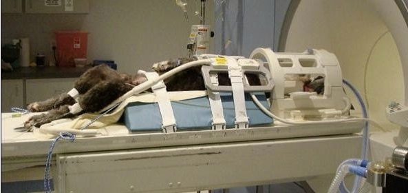
Veterinary MRI Safety: Heating & Patient Burn Prevention
MRI (Magnetic Resonance Imaging) is considered to be a safe medical imaging technique. It does not use ionizing radiation like X-rays or CT scans, which can potentially damage cells or DNA. Instead, MRI uses strong magnetic fields and radio waves to generate images of the inside of the body. In veterinary medicine we must anesthetize the patient when scanning so that they remain completely still and properly positioned for the scan. Due to the small size of most veterinary patients, hypothermia is much more common during general anesthesia, however, industry wide we are receiving increased requests for information on patient heating and burn.
Patient burn and heating in the context of MRI (Magnetic Resonance Imaging) refer to two different phenomena that can occur during the imaging process. But what are the differences, why are they concerns, and how do we prevent them?
Patient heating in MRI (Magnetic Resonance Imaging) can occur due to a phenomenon called “RF (Radiofrequency) heating.” RF heating happens when radiofrequency energy is absorbed by the patient’s tissues during an MRI scan. This can lead to a rise in the patient’s core body temperature, and while generally not a problem in human medicine, can be serious in veterinary medicine if cooling measures are not taken immediately.
Some heating prevention measures include:
- Patient Monitoring: Dogs and cats are under general anesthesia while they are scanned, and anesthetic drugs blunt patients’ ability to regulate their own body temperature. They have a reduced ability to detect increases in core body temperature, and therefore their compensatory responses to hyperthermia, such as panting, may be delayed or incomplete. Constant monitoring of the patient’s vital signs, including heart rate, respiratory rate, and end-tidal CO2, is essential so that abnormal changes can be promptly addressed. For this reason AnimalScan recommends utilizing the proper MRI conditional ancillary equipment, such as anesthesia monitors outfitted with the proper pediatric readings and connectors.
- Coil Selection: Choose appropriate radiofrequency coils based on the patient’s size and anatomy. Using coils that are too large for the patient can lead to excessive heating due to interactions with the magnetic field.
- Limiting Scan Time: Longer MRI scan times can increase the risk of heating. Whenever possible, use shorter scan sequences to minimize the duration of exposure and plan ahead for events such as adding contrast or repositioning the patient.
- Appropriate Sequences: Choose MRI sequences that minimize the amount of energy deposited into the patient’s body. Some sequences are known to cause more heating than others. Confirm that your radiologist or MRI technologist has first hand experience working with your specific machine and has developed protocols aimed at minimizing scan times and maximizing diagnostic accuracy. Working with an experienced veterinary radiologist and applications specialist will greatly reduce your chances of causing a heating event.
- Testing and Validation: Regularly test and validate the MRI system’s safety measures to ensure they are functioning as intended. This includes assessing the accuracy of temperature monitoring and emergency shutdown systems.
- Expertise and Training: Ensure that the veterinary team operating the MRI equipment is properly trained in animal safety during MRI procedures. They should be knowledgeable about potential risks and how to mitigate them.
- Emergency Procedures: Have clear protocols in place for what to do in case of patient heating, including stopping the scan and initiating cooling measures.
Patient burn occurs when metallic objects or implants in a patient’s body increase in temperature due to the RF energy used during the scan. When a metallic object or implant is exposed to the RF energy in an MRI machine, it can absorb some of that energy and convert it into heat. This can lead to a localized increase in temperature in the vicinity of the metal. Significant heating can potentially be harmful, particularly if the metal object is large, highly conductive, or in close proximity to sensitive tissues. This is why it’s important to assess the safety of any metallic implants before an MRI as well as all medical supplies and equipment that may come in contact with the patient during the scan. Some examples of this include:
- Vet wrap, which may contain metallic fibers which cause burn during a scan.
- ECG and pulse ox wireless modules
- BB Shot embedded in a patient’s skin
- Implants
Below are some of the measures that can be taken to prevent these uncomfortable and potentially harmful conditions from occuring in our patients.
- Pre-Scan Assessment: Before the MRI, assess the patient’s condition thoroughly. Ensure they are not carrying any metallic implants, devices, or objects that could heat up or cause harm during the procedure. Preprocedural radiographs can be helpful in identifying these objects.
- Patient Positioning: Properly position the patient to minimize contact between metallic objects and the patient’s body. Make sure there are no metallic collars, tags, or accessories on the animal. Position ECG and pulse oximeter wireless modules away from the patient and outside of the coil.
- Consultation with Veterinary Radiologists and Physicists: Collaborate with experienced veterinary radiologists and medical physicists to ensure that the MRI protocols and safety measures are appropriate for the specific patient and procedure. It is extremely important that your radiologist have expertise in veterinary MRI.
Both burn and heating are concerns in MRI safety, and precautions are taken to ensure patient comfort and safety during the imaging process. Remember that each patient is unique, and the precautions taken should be tailored to their individual needs. Regular review of safety protocols and staying up-to-date with the latest research in veterinary MRI safety is crucial for maintaining patient well-being.


Leave a Reply